


Please enter the answer below before you can view the full text.
8+4=
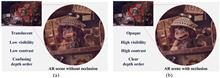
We propose a near-eye display optics system that supports three-dimensional mutual occlusion. By exploiting the polarization-control properties of a phase-only liquid crystal on silicon (LCoS), we achieve real see-through scene masking as well as virtual digital scene imaging using a single LCoS. Dynamic depth control of the real scene mask and virtual digital image is also achieved by using a focus tunable lens (FTL) pair of opposite curvatures. The proposed configuration using a single LCoS and opposite curvature FTL pair enables the self-alignment of the mask and image at an arbitrary depth without distorting the see-through view of the real scene. We verified the feasibility of the proposed optics using two optical benchtop setups: one with two off-the-shelf FTLs for continuous depth control, and the other with a single Pancharatnam–Berry phase-type FTL for the improved form factor.
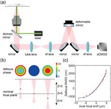
Single-molecule localization microscopy (SMLM) enables three-dimensional (3D) investigation of nanoscale structures in biological samples, offering unique insights into their organization. However, traditional 3D super-resolution microscopy using high numerical aperture (NA) objectives is limited by imaging depth of field (DOF), restricting their practical application to relatively thin biological samples. Here, we developed a unified solution for thick sample super-resolution imaging using a deformable mirror (DM) which served for fast remote focusing, optimized point spread function (PSF) engineering, and accurate aberration correction. By effectively correcting the system aberrations introduced during remote focusing and sample aberrations at different imaging depths, we achieved high-accuracy, large DOF imaging (∼8 μm) of the whole-cell organelles [i.e., nuclear pore complex (NPC), microtubules, and mitochondria] with a nearly uniform resolution of approximately 35 nm across the entire cellular volume.
The generation of speckle patterns via random matrices, statistical definitions, or apertures may not always result in optimal outcomes. Issues such as correlation fluctuations in low ensemble numbers and diffraction in long-distance propagation can arise. Instead of improving results of specific applications, our solution is catching deep correlations of patterns with the framework, Speckle-Net, which is fundamental and universally applicable to various systems. We demonstrate this in computational ghost imaging (CGI) and structured illumination microscopy (SIM). In CGI with extremely low ensemble number, it customizes correlation width and minimizes correlation fluctuations in illuminating patterns to achieve higher-quality images. It also creates non-Rayleigh nondiffracting speckle patterns only through a phase mask modulation, which overcomes the power loss in the traditional ring-aperture method. Our approach provides new insights into the nontrivial speckle patterns and has great potential for a variety of applications including dynamic SIM, X-ray and photo-acoustic imaging, and disorder physics.
Structured illumination microscopy (SIM) has been widely applied to investigate intricate biological dynamics due to its outstanding super-resolution imaging speed. Incorporating compressive sensing into SIM brings the possibility to further improve the super-resolution imaging speed. Nevertheless, the recovery of the super-resolution information from the compressed measurement remains challenging in experiments. Here, we report structured illumination microscopy with complementary encoding-based compressive imaging (CECI-SIM) to realize faster super-resolution imaging. Compared to the nine measurements to obtain a super-resolution image in a conventional SIM, CECI-SIM can achieve a super-resolution image by three measurements; therefore, a threefold improvement in the imaging speed can be achieved. This faster imaging ability in CECI-SIM is experimentally verified by observing tubulin and actin in mouse embryonic fibroblast cells. This work provides a feasible solution for high-speed super-resolution imaging, which would bring significant applications in biomedical research.
With the swift advancement of neural networks and their expanding applications in many fields, optical neural networks have gradually become a feasible alternative to electrical neural networks due to their parallelism, high speed, low latency, and power consumption. Nonetheless, optical nonlinearity is hard to realize in free-space optics, which restricts the potential of the architecture. To harness the benefits of optical parallelism while ensuring compatibility with natural light scenes, it becomes essential to implement two-dimensional spatial nonlinearity within an incoherent light environment. Here, we demonstrate a lensless opto-electrical neural network that incorporates optical nonlinearity, capable of performing convolution calculations and achieving nonlinear activation via a quantum dot film, all without an external power supply. Through simulation and experiments, the proposed nonlinear system can enhance the accuracy of image classification tasks, yielding a maximum improvement of 5.88% over linear models. The scheme shows a facile implementation of passive incoherent two-dimensional nonlinearities, paving the way for the applications of multilayer incoherent optical neural networks in the future.
The exact physical modeling for scattered light modulation is critical in phototherapy, biomedical imaging, and free-space optical communications. In particular, the angular spectrum modeling of scattered light has attracted considerable attention, but the existing angular spectrum models neglect the polarization of photons, degrading their performance. Here, we propose a full-polarization angular spectrum model (fpASM) to take the polarization into account. This model involves a combination of the optical field changes and free-space angular spectrum diffraction, and enables an investigation of the influence of polarization-related factors on the performance of scattered light modulation. By establishing the relationship between various model parameters and macroscopic scattering properties, our model can effectively characterize various depolarization conditions. As a demonstration, we apply the model in the time-reversal data transmission and anti-scattering light focusing. Our method allows the analysis of various depolarization scattering events and benefits applications related to scattered light modulation.
Optical aberrations degrade the performance of fluorescence microscopy. Conventional adaptive optics (AO) leverages specific devices, such as the Shack–Hartmann wavefront sensor and deformable mirror, to measure and correct optical aberrations. However, conventional AO requires either additional hardware or a more complicated imaging procedure, resulting in higher cost or a lower acquisition speed. In this study, we proposed a novel space-frequency encoding network (SFE-Net) that can directly estimate the aberrated point spread functions (PSFs) from biological images, enabling fast optical aberration estimation with high accuracy without engaging extra optics and image acquisition. We showed that with the estimated PSFs, the optical aberration can be computationally removed by the deconvolution algorithm. Furthermore, to fully exploit the benefits of SFE-Net, we incorporated the estimated PSF with neural network architecture design to devise an aberration-aware deep-learning super-resolution model, dubbed SFT-DFCAN. We demonstrated that the combination of SFE-Net and SFT-DFCAN enables instant digital AO and optical aberration-aware super-resolution reconstruction for live-cell imaging.
Biodynamical processes, especially in system biology, that occur far apart in space may be highly correlated. To study such biodynamics, simultaneous imaging over a large span at high spatio-temporal resolutions is highly desired. For example, large-scale recording of neural network activities over various brain regions is indispensable in neuroscience. However, limited by the field-of-view (FoV) of conventional microscopes, simultaneous recording of laterally distant regions at high spatio-temporal resolutions is highly challenging. Here, we propose to extend the distance of simultaneous recording regions with a custom micro-mirror unit, taking advantage of the long working distance of the objective and spatio-temporal multiplexing. We demonstrate simultaneous dual-region two-photon imaging, spanning as large as 9 mm, which is 4 times larger than the nominal FoV of the objective. We verify the system performance in in vivo imaging of neural activities and vascular dilations, simultaneously, at two regions in mouse brains as well as in spinal cords, respectively. The adoption of our proposed scheme will promote the study of systematic biology, such as system neuroscience and system immunology.
The pyramid wavefront sensor (PWFS) can provide the sensitivity needed for demanding adaptive optics applications, such as imaging exoplanets using the future extremely large telescopes of over 30 m of diameter (D). However, its exquisite sensitivity has a limited linear range of operation, or dynamic range, although it can be extended through the use of beam modulation—despite sacrificing sensitivity and requiring additional optical hardware. Inspired by artificial intelligence techniques, this work proposes to train an optical layer—comprising a passive diffractive element placed at a conjugated Fourier plane of the pyramid prism—to boost the linear response of the pyramid sensor without the need for cumbersome modulation. We develop an end-2-end simulation to train the diffractive element, which acts as an optical preconditioner to the traditional least-square modal phase estimation process. Simulation results with a large range of turbulence conditions show a noticeable improvement in the aberration estimation performance equivalent to over 3λ/D of modulation when using the optically preconditioned deep PWFS (DPWFS). Experimental results validate the advantages of using the designed optical layer, where the DPWFS can pair the performance of a traditional PWFS with 2λ/D of modulation. Designing and adding an optical preconditioner to the PWFS is just the tip of the iceberg, since the proposed deep optics methodology can be used for the design of a completely new generation of wavefront sensors that can better fit the demands of sophisticated adaptive optics applications such as ground-to-space and underwater optical communications and imaging through scattering media.
Light-sheet fluorescence microscopy (LSFM) has played an important role in bio-imaging due to its advantages of high photon efficiency, fast speed, and long-term imaging capabilities. The perpendicular layout between LSFM excitation and detection often limits the 3D resolutions as well as their isotropy. Here, we report on a reflective type light-sheet microscope with a mini-prism used as an optical path reflector. The conventional high NA objectives can be used both in excitation and detection with this design. Isotropic resolutions in 3D down to 300 nm could be achieved without deconvolution. The proposed method also enables easy transform of a conventional fluorescence microscope to high performance light-sheet microscopy.
Ultraviolet (UV) imaging enables a diverse array of applications, such as material composition analysis, biological fluorescence imaging, and detecting defects in semiconductor manufacturing. However, scientific-grade UV cameras with high quantum efficiency are expensive and include complex thermoelectric cooling systems. Here, we demonstrate a UV computational ghost imaging (UV-CGI) method to provide a cost-effective UV imaging and detection strategy. By applying spatial–temporal illumination patterns and using a 325 nm laser source, a single-pixel detector is enough to reconstruct the images of objects. We use UV-CGI to distinguish four UV-sensitive sunscreen areas with different densities on a sample. Furthermore, we demonstrate dark-field UV-CGI in both transmission and reflection schemes. By only collecting the scattered light from objects, we can detect the edges of pure phase objects and small scratches on a compact disc. Our results showcase a feasible low-cost solution for nondestructive UV imaging and detection. By combining it with other imaging techniques, such as hyperspectral imaging or time-resolved imaging, a compact and versatile UV computational imaging platform may be realized for future applications.
Non-line-of-sight (NLOS) imaging is a challenging task aimed at reconstructing objects outside the direct view of the observer. Nevertheless, traditional NLOS imaging methods typically rely on intricate and costly equipment to scan and sample the hidden object. These methods often suffer from restricted imaging resolution and require high system stability. Herein, we propose a single-shot high-resolution NLOS imaging method via chromato-axial differential correlography, which adopts low-cost continuous-wave lasers and a conventional camera. By leveraging the uncorrelated laser speckle patterns along the chromato-axis, this method can reconstruct hidden objects of diverse complexity using only one exposure measurement. The achieved background stability through single-shot acquisition, along with the inherent information redundancy in the chromato-axial differential speckles, enhances the robustness of the system against vibration and colored stain interference. This approach overcomes the limitations of conventional methods by simplifying the sampling process, improving system stability, and achieving enhanced imaging resolution using available equipment. This work serves as a valuable reference for the real-time development and practical implementation of NLOS imaging.
Using freeform optical surfaces in lens design can lead to much higher system specifications and performance while significantly reducing volume and weight. However, because of the complexity of freeform surfaces, freeform optical design using traditional methods requires extensive human effort and sufficient design experience, while other design methods have limitations in design efficiency, simplicity, and versatility. Deep learning can solve these issues by summarizing design knowledge and applying it to design tasks with different system and structure parameters. We propose a deep-learning framework for designing freeform imaging systems. We generate the data set automatically using a combined sequential and random system evolution method. We combine supervised learning and unsupervised learning to train the network so that it has good generalization ability for a wide range of system and structure parameter values. The generated network FreeformNet enables fast generation (less than 0.003 s per system) of multiple-solution systems after we input the design requirements, including the system and structure parameters. We can filter and sort solutions based on a given criterion and use them as good starting points for quick final optimization (several seconds for systems with small or moderate field-of-view in general). The proposed framework presents a revolutionary approach to the lens design of freeform or generalized imaging systems, thus significantly reducing the time and effort expended on optical design.
In this paper, we propose a real-time incoherent digital holographic (IDH) recording system free from bias and twin-image noises. A motionless three-step polarization-encoded phase-shifter operating at 99 Hz is realized with two electrically controllable birefringence-mode liquid crystal cells operating in tandem with a geometric phase lens and polarizers. Based on the proposed optical configuration, a coaxial straight-line self-interference IDH recording system is devised. Notably, the elimination of bias and twin-image noise from three phase-shifted images is demonstrated as a proof of concept. Moreover, complex-valued holographic video acquisitions with a resolution greater than 20 megapixels are demonstrated, with an effective acquisition frequency of 33 Hz.
Imaging through scattering media is valuable for many areas, such as biomedicine and communication. Recent progress enabled by deep learning (DL) has shown superiority especially in the model generalization. However, there is a lack of research to physically reveal the origin or define the boundary for such model scalability, which is important for utilizing DL approaches for scalable imaging despite scattering with high confidence. In this paper, we find the amount of the ballistic light component in the output field is the prerequisite for endowing a DL model with generalization capability by using a “one-to-all” training strategy, which offers a physical meaning invariance among the multisource data. The findings are supported by both experimental and simulated tests in which the roles of scattered and ballistic components are revealed in contributing to the origin and physical boundary of the model scalability. Experimentally, the generalization performance of the network is enhanced by increasing the portion of ballistic photons in detection. The mechanism understanding and practical guidance by our research are beneficial for developing DL methods for descattering with high adaptivity.
Rather than focusing on a focal spot, aberrated wavefields spread out over a region. As a wave phenomenon, optical aberrations are analyzed in terms of waves propagating in the 3D space. In this work, we report the observation of 2D longitudinal aberrated wavefields. This observation can be visualized by mapping the intensity distributions of surface plasmon polaritons (SPPs) that propagate on a metal/air interface using leakage radiation microscopy. The orientation of the SPP beam is tweaked by tilting and translating the system to mimic aberrated beams, presenting known Seidel terms: defocus, spherical, coma, and tilt aberration. This approach allows the examination of the longitudinal evolution of aberrated beams in a visual and rapid manner, in contrast to more complicated post-processing reconstructions.
Förster resonance energy transfer (FRET) microscopy provides unique insight into the functionality of biological systems via imaging the spatiotemporal interactions and functional state of proteins. Distinguishing FRET signals from sub-diffraction regions requires super-resolution (SR) FRET imaging, yet is challenging to achieve from living cells. Here, we present an SR FRET method named SIM-FRET that combines SR structured illumination microscopy (SIM) imaging and acceptor sensitized emission FRET imaging for live-cell quantitative SR FRET imaging. Leveraging the robust co-localization prior of donor and accepter during FRET, we devised a mask filtering approach to mitigate the impact of SIM reconstruction artifacts on quantitative FRET analysis. Compared to wide-field FRET imaging, SIM-FRET provides nearly twofold spatial resolution enhancement of FRET imaging at sub-second timescales and maintains the advantages of quantitative FRET analysis in vivo. We validate the resolution enhancement and quantitative analysis fidelity of SIM-FRET signals in both simulated FRET models and live-cell FRET-standard construct samples. Our method reveals the intricate structure of FRET signals, which are commonly distorted in conventional wide-field FRET imaging.
Micro-LEDs are one of the most promising candidates for next-generation displays, yet they are inconvenienced by the efficiency reduction induced by the sidewall defects when pursuing further scaled-down device dimensions. We have systematically investigated both the size and temporal dependence of micro-LEDs. Micro-LED arrays with a mesa size ranging from 7 to 100 μm were prepared for display purposes. The luminance and external quantum efficiency (EQE) were measured and discussed. Surprisingly, micro-LED arrays with a smaller mesa size exhibit a higher EQE under 100 ns pulse duration operation when compared with longer pulse duration operations. Under certain short-pulsed excitation, a 7×7 μm2 micro-LED array even exhibits a >20% higher EQE as compared to the direct current (DC) or the long duration pulse operation condition. We thus concluded that the notorious efficiency reduction induced by sidewall defects in small-sized micro-LED arrays could be significantly reduced by applying short-pulse voltages.
Quantitative phase imaging (QPI) by differential phase contrast (DPC) with partially coherent illumination provides speckle-free imaging and lateral resolution beyond the coherent diffraction limit, demonstrating great potential in biomedical imaging applications. Generally, DPC employs weak object approximation to linearize the phase-to-intensity image formation, simplifying the solution to the phase retrieval as a two-dimensional deconvolution with the corresponding phase transfer function. Despite its widespread adoption, weak object approximation still lacks a precise and clear definition, suggesting that the accuracy of the QPI results, especially for samples with large phase values, is yet to be verified. In this paper, we analyze the weak object approximation condition quantitatively and explicitly give its strict definition that is applicable to arbitrary samples and illumination apertures. Furthermore, an iterative deconvolution QPI technique based on pseudo-weak object approximation is proposed to overcome the difficulty of applying DPC to large-phase samples without additional data acquisition. Experiments with standard microlens arrays and MCF-7 cells demonstrated that the proposed method can effectively extend DPC beyond weak object approximation to high-precision three-dimensional morphological characterization of large-phase technical and biological samples.
Computed tomography imaging spectrometry (CTIS) is a snapshot spectral imaging technique that relies on a limited number of projections of the target data cube (2D spatial and 1D spectral), which can be reconstructed via a delicate tomographic reconstruction algorithm. However, the restricted angle difference between the projections and the space division multiplexing of the projections make the reconstruction suffer from severe artifacts as well as a low spatial resolution. In this paper, we demonstrate super-resolution computed tomography imaging spectrometry (SRCTIS) by assimilating the information obtained by a conventional CTIS system and a regular RGB camera, which has a higher pixel resolution. To improve the reconstruction accuracy of CTIS, the unique information provided by the zero-order diffraction of the target scene is used as a guidance image for filtering to better preserve the edges and reduce artifacts. The recovered multispectral image is then mapped onto the RGB image according to camera calibration. Finally, based on the spectral and the spatial continuities of the target scene, the multispectral information obtained from CTIS is propagated to each pixel of the RGB image to enhance its spectral resolution, resulting in SRCTIS. Both stimulative studies and proof-of-concept experiments were then conducted, and the results quantified by key metrics, such as structural similarity index measurement and spectral angle mapping have suggested that the developed method cannot only suppress the reconstruction artifacts, but also simultaneously achieve high spatial and spectral resolutions.
Polarimetric imaging provides valuable insights into the polarization state of light interacting with a sample. It can infer crucial birefringence properties of specimens without using labels, thereby facilitating the diagnosis of diseases such as cancer and osteoarthritis. In this study, we present a novel polarimetric coded ptychography (pol-CP) approach that enables high-resolution, high-throughput gigapixel birefringence imaging on a chip. Our platform deviates from traditional lens-based systems by employing an integrated polarimetric coded sensor for lensless coherent diffraction imaging. Utilizing Jones calculus, we quantitatively determine the birefringence retardance and orientation information of biospecimens from the recovered images. Our portable pol-CP prototype can resolve the 435 nm linewidth on the resolution target, and the imaging field of view for a single acquisition is limited only by the detector size of 41 mm2. The prototype allows for the acquisition of gigapixel birefringence images with a 180 mm2 field of view in ∼3.5 min, a performance that rivals high-end whole slide scanner but at a small fraction of the cost. To demonstrate its biomedical applications, we perform high-throughput imaging of malaria-infected blood smears, locating parasites using birefringence contrast. We also generate birefringence maps of label-free thyroid smears to identify thyroid follicles. Notably, the recovered birefringence maps emphasize the same regions as autofluorescence images, underscoring the potential for rapid on-site evaluation of label-free biopsies. Our approach provides a turnkey and portable solution for lensless polarimetric analysis on a chip, with promising applications in disease diagnosis, crystal screening, and label-free chemical imaging, particularly in resource-constrained environments.
Optical-resolution photoacoustic microscopy (OR-PAM) is capable of observing the distribution of optical absorbers inside bio-tissues with a high spatial resolution of micrometers. Unfortunately, due to the employment of a tight optical focus, it suffers from a limited depth of field (DOF), making it challenging to achieve high-resolution imaging of targets with arbitrary surfaces. Here, we propose a high spatiotemporal adaptive photoacoustic focusing mechanism through integrating a high-speed optical focuser, a time-of-flight contour deriving algorithm, and the rotary-scanning photoacoustic microscopy. The developed system, named high-speed adaptive photoacoustic microscopy (HA-PAM), features an ultrashort focus-shifting time of 5 ms and an enlarged DOF of up to 5 mm. With the assistance of the proposed mechanism, we can achieve a homogeneous lateral resolution of 6 μm over a 10 mm circular imaging domain within 5 s. We demonstrate the advantages of HA-PAM through imaging phantoms with curved surfaces, subcutaneous tumor-bearing mice, resected rabbit kidneys, and pulsating mouse brains. The imaging results suggest that this approach provides a high and consistent spatial resolution for imaging bio-tissues with arbitrary surfaces without sacrificing the imaging speed, and has the potential to extend the fundamental and clinical applications of OR-PAM.
Fiber scanners are portable and miniaturized laser scanning devices used for a wide range of applications, such as endoscopic probes for biomedical imaging. However, in order to achieve different resonant frequencies for 2D actuation, existing fiber scanners have complex actuation mechanisms and structures, resulting in being an obstacle for endoscopic imaging. By exploiting the intrinsic difference in bending stiffness of non-symmetrical fibers, we present the most simplified fiber scanner to date, containing only a single piezoelectric bimorph and a single non-symmetrical fiber with a 1D actuator for 2D laser scanning. 5-fps (frames per second) Lissajous scan is achieved with a scanning range of >300 μm and a driving voltage of ≤10Vpp. The ultra simplified structure of the fiber scanner enables a miniaturized optical probe with a diameter of 1.9 mm, and image quality comparable to that of commercial microscopes. Taking advantage of its ease of manufacture and low cost, the fiber scanner offers a transformative way forward for disposable endoscopic probes that avoid the risk of cross infection during endoscopic inspections.
Photoacoustic endomicroscopy combined with ultrasound (PAEM-US) has been a long-standing expectation for gastrointestinal tumor examination. Here, we introduce a prototype disposable PAEM-US catheter and corresponding power interface unit, featuring catheter switchability, self-internal three-dimensional scanning, and system repeatability for gastrointestinal endoscopy. By utilizing high-fluence relays, cascade insertion loss of the optical waveguide is minimized to 0.6 dB with a high performance of power resistance, and a focus-customizable acousto-optic coaxial probe is designed for high-sensitivity optical-resolution photoacoustic imaging. Imaging capability was demonstrated with in vivo anatomical imaging at 30 frames per second. Imaging results showed co-registered microscopic visualization of the microvascular and stratification of the rat colorectum with lateral resolution of 18 μm and axial resolution of 63 μm, holding great potential in the clinical detection of gastrointestinal diseases.
Artificial neural networks have shown great proficiency in transforming low-resolution microscopic images into high-resolution images. However, training data remains a challenge, as large-scale open-source databases of microscopic images are rare, particularly 3D data. Moreover, the long training times and the need for expensive computational resources have become a burden to the research community. We introduced a deep-learning-based self-supervised volumetric imaging approach, which we termed “Self-Vision.” The self-supervised approach requires no training data, apart from the input image itself. The lightweight network takes just minutes to train and has demonstrated resolution-enhancing power on par with or better than that of a number of recent microscopy-based models. Moreover, the high throughput power of the network enables large image inference with less postprocessing, facilitating a large field-of-view (2.45 mm×2.45 mm) using a home-built two-photon microscopy system. Self-Vision can recover images from fourfold undersampled inputs in the lateral and axial dimensions, dramatically reducing the acquisition time. Self-Vision facilitates the use of a deep neural network for 3D microscopy imaging, easing the demanding process of image acquisition and network training for current resolution-enhancing networks.
Micro-endoscopes are widely used for detecting and visualizing hard-to-reach areas of the human body and for in vivo observation of animals. A micro-endoscope that can realize 3D imaging at the camera framerate could benefit various clinical and biological applications. In this work, we report the development of a compact light-field micro-endoscope (LFME) that can obtain snapshot 3D fluorescence imaging, by jointly using a single-mode fiber bundle and a small-size light-field configuration. To demonstrate the real imaging performance of our method, we put a resolution chart in different z positions and capture the z-stack images successively for reconstruction, achieving 333-μm-diameter field of view, 24 μm optimal depth of field, and up to 3.91 μm spatial resolution near the focal plane. We also test our method on a human skin tissue section and HeLa cells. Our LFME prototype provides epi-fluorescence imaging ability with a relatively small (2-mm-diameter) imaging probe, making it suitable for in vivo detection of brain activity and gastrointestinal diseases of animals.
Single-pixel imaging (SPI) can capture 2D images of the target with only a nonpixelated detector, showing promising application potential in nonvisible spectral imaging, low-photon imaging, lidar, and other extreme imaging fields. However, the imaging mechanism of traditional SPI makes it difficult to achieve high imaging speed, which is a primary barrier for its widespread application. To address this issue, in this work, we propose and demonstrate a novel high-speed 2D and 3D imaging scheme based on traditional SPI, termed time-resolved single-pixel imaging (TRSPI). Previous SPI works mainly utilize correlation between a stable target and iterative illumination masks to reconstruct a single image. In TRSPI, by further exploiting correlation information between a dynamic scene and every static mask, we can reconstruct a series of time-varying images of the dynamic scene, given the dynamic scene is repetitive or reproducible. Experimentally, we conducted 2D and 3D imaging on a rotating chopper with a speed of 4800 revolutions per minute (rpm), and imaging speeds up to 2,000,000 fps. It is believed that this technology not only opens up a novel application direction for SPI, but also will provide a powerful solution for high-speed imaging.
Nitrogen vacancy diamonds have emerged as sensitive solid-state magnetic field sensors capable of producing diffraction limited and sub-diffraction field images. Here, for the first time, to our knowledge, we extend those measurements to high-speed imaging, which can be readily applied to analyze currents and magnetic field dynamics in circuits on a microscopic scale. To overcome detector acquisition rate limitations, we designed an optical streaking nitrogen vacancy microscope to acquire two-dimensional spatiotemporal kymograms. We demonstrate magnetic field wave imaging with micro-scale spatial extent and ∼400 μs temporal resolution. In validating this system, we detected magnetic fields down to 10 μT for 40 Hz magnetic fields using single-shot imaging and captured the spatial transit of an electromagnetic needle at streak rates as high as 110 μm/ms. This design has the capability to be readily extended to full 3D video acquisition by utilizing compressed sensing techniques and a potential for further improvement of spatial resolution, acquisition speed, and sensitivity. The device opens opportunities to many potential applications where transient magnetic events can be isolated to a single spatial axis, such as acquiring spatially propagating action potentials for brain imaging and remotely interrogating integrated circuits.
Imaging ultrafast processes in femtosecond (fs) laser–material interactions such as fs laser ablation is very important to understand the physical mechanisms involved. To achieve this goal with high resolutions in both spatial and temporal domains, a combination of optical pump–probe microscopy and structured illumination microscopy can be a promising approach, but suffers from the multiple-frame method with a phase shift that is inapplicable to irreversible ultrafast processes such as ablation. Here, we propose and build a wide-field single-probe structured light microscopy (SPSLM) to image the ultrafast three-dimensional topography evolution induced by fs lasers, where only a single imaging frame with a single structured probe pulse is required for topography reconstruction, benefiting from Fourier transform profilometry. The second harmonic of the fs laser is used as the structured probe light to improve spatial lateral resolution into the subwavelength region of ∼478 nm, and the spatial axial and temporal resolutions are estimated to be ∼22 nm and ∼256 fs, respectively. With SPSLM, we successfully image the ultrafast topography evolution of a silicon wafer surface impacted by single and multiple fs pulses. The variable formation and evolution of the laser induced periodic surface structures during an ultrashort time are visualized and analyzed. We believe that SPSLM will be a significant approach for revealing and understanding various ultrafast dynamics, especially in fs laser ablation and material science.
To assess the performance of adaptive optics and predict an optimal wavefront correction, we built a wavefront reconstructor with a damped transpose matrix of the influence function. Using an integral control strategy, we tested this reconstructor with four deformable mirrors in an experimental system, an adaptive optics scanning laser ophthalmoscope, and an adaptive optics near-confocal ophthalmoscope. Testing results proved that this reconstructor could ensure a stable and precise correction for wavefront aberration compared to a conventional optimal reconstructor formed by the inverse matrix of the influence function. This method may provide a helpful tool for testing, evaluating, and optimizing adaptive optics systems.
Optical imaging through scattering media has long been a challenge. Many approaches have been developed for focusing light or imaging objects through scattering media, but usually, they are either invasive, limited to stationary or slow-moving media, or require high-resolution cameras and complex algorithms to retrieve the images. By utilizing spatial–temporal encoded patterns (STEPs), we introduce a technique for the computation of imaging that overcomes these restrictions. With a single-pixel photodetector, we demonstrate non-invasive imaging through scattering media. This technique is insensitive to the motion of the media. Furthermore, we demonstrate that our image reconstruction algorithm is much more efficient than correlation-based algorithms for single-pixel imaging, which may allow fast imaging for applications with limited computing resources.
Conventional ptychography translates an object through a localized probe beam to widen the field of view in real space. Fourier ptychography translates the object spectrum through a pupil aperture to expand the Fourier bandwidth in reciprocal space. Here we report an imaging modality, termed synthetic aperture ptychography (SAP), to get the best of both techniques. In SAP, we illuminate a stationary object using an extended plane wave and translate a coded image sensor at the far field for data acquisition. The coded layer attached on the sensor modulates the object exit waves and serves as an effective ptychographic probe for phase retrieval. The sensor translation process in SAP synthesizes a large complex-valued wavefront at the intermediate aperture plane. By propagating this wavefront back to the object plane, we can widen the field of view in real space and expand the Fourier bandwidth in reciprocal space simultaneously. We validate the SAP approach with transmission targets and reflection silicon microchips. A 20-mm aperture was synthesized using a 5-mm sensor, achieving a fourfold gain in resolution and 16-fold gain in field of view for object recovery. In addition, the thin sample requirement in ptychography is no longer required in SAP. One can digitally propagate the recovered exit wave to any axial position for post-acquisition refocusing. The SAP scheme offers a solution for far-field sub-diffraction imaging without using lenses. It can be adopted in coherent diffraction imaging setups with radiation sources from visible light, extreme ultraviolet, and X-ray, to electron.
A remarkable feature of Alvarez lenses is that a wide focal length tuning range can be achieved using lateral displacement rather than commonly used axial translation, thus, reducing the overall length of varifocal imaging systems. Here, we present novel lens elements based on Alvarez lenses actuated by a dielectric elastomer (DE). The proposed lens elements are composed of the varifocal component and the scanning component. Based on the proposed lens elements, an imaging system is built to realize ultra-wide varifocal imaging with a selectable region of interest. The lens elements have a variable focus function based on an Alvarez lens structure and a DE actuator and a scanning function based on the DE-based four-quadrant actuators. The large deformation generated by the DE actuators permits the lateral displacement of the Alvarez lenses up to 1.145 mm. The focal length variation of the proposed varifocal component is up to 30.5 times, where the maximum focal length is 181 mm and the minimum focal length is 5.94 mm. The rise and fall times of the varifocal component are 160 ms and 295 ms, respectively. By applying different voltages on four-quadrant actuators, the scanning component allows the varifocal component to move in different directions and endows the varifocal component with a selectable region of interest imaging capability. The scanning range of the scanning component is 17.57°. The imaging resolution of the imaging system is approximately 181 lp/mm. The system developed in the current study has the potential to be used in consumer electronics, endoscopy, and microscopy in the future.
Stereoscopic microscopy is a promising technology to obtain three-dimensional microscopic images. Such microscopes are based on the parallax effect, and as such require two lenses to focus at two different points. Geometrical constraints, however, restrict their numerical apertures to about 0.2, thus limiting the system’s resolution. Higher numerical apertures (∼0.35) can be achieved with designs using only one bulk lens, but such systems are ∼10 times more costly than the conventional ones. Thus, there is a pressing need for alternative solutions to improve the resolution of stereoscopic systems. Here, we show that high-resolution and low-cost stereoscopic systems can be obtained using birefringent single-layer metalenses. We design and fabricate a birefringent metalens operating at 532 nm with a numerical aperture as high as 0.4. The metalens is then used to demonstrate high-resolution stereoscopic imaging of biological samples. The microscopic images are further displayed and perceived vividly in an autostereoscopic display. Our demonstration paves the way to a new strategy to achieve high-resolution and low-cost stereoscopic microscopes.
Structured illumination microscopy (SIM) is an advanced microscope system that provides superresolution capability with excellent imaging speed, which has become a practical tool for live-cell imaging. However, the bulky size is blocking the application of SIM in wider study fields and scenarios. Here, we developed a miniaturized SIM (Mini SIM) system that provided periodic illumination using a diffractive optical element (DOE) for the first time. This optimized phase-only DOE generated the two-dimensional sinusoidal illumination by optical Fourier transform with an illuminating objective lens, which substantially simplified and miniaturized the illumination system. We built up a Mini SIM prototype and demonstrated lateral superresolution imaging of fluorescence beads and A549 cell slides. The proposed Mini SIM greatly simplifies the experimental setup and may lead to important applications in bio-imaging.
Polarization optical imaging can be used to characterize anisotropy in biological tissue microstructures and has been demonstrated to be a powerful tool for clinical diagnosis. However, the approach is limited by an inability to image targets deeper than ∼1 mm due to strong optical scattering in biological tissues. As such, we propose a novel polarization microwave-induced thermoacoustic imaging (P-MTAI) technique to noninvasively detect variations in deep tissue by exploiting the thermoacoustic signals induced by four pulsed microwaves of varying polarization orientations. The proposed P-MTAI method overcomes the penetration limits of conventional polarization optical imaging and provides submillimeter resolution over depths of several centimeters. As part of the paper, the structural characteristics of tissues were quantified using a new parameter, the degree of microwave absorption anisotropy. P-MTAI was also applied to the noninvasive detection of morphological changes in cardiomyocytes as they transitioned from ordered to disordered states, providing a potential indication of myocardial infarction.
Fourier light field microscopy (FLFM) shows great potential in high-speed volumetric imaging of biodynamics. However, due to the inherent disadvantage of wide-field illumination, it suffers from intense background, arising from out of the depth-of-field signal and tissue scattered noise. The background will not only deteriorate the image contrast, making quantitative measurement difficult, but also introduce artifacts, especially in functional imaging of the neuronal network activity in vivo. Here, we propose the robust Fourier light field microscopy (RFLFM), which suppresses the background in FLFM by introducing structured illumination and computational reconstruction based on HiLo. The superior performance of RFLFM is verified by volumetric imaging of biological dynamics in larval zebrafish and mouse in vivo, at a volumetric imaging rate up to 33.3 Hz. The statistical results show that the fluorescence background can be significantly depressed, with the signal-to-background ratio improved by orders of magnitude and the whole image contrast improved by as much as ∼10.4 times. Moreover, we stress that, in functional imaging of neuronal network activity in turbid brain tissues, our system can avoid artifacts resulting from background fluctuations, while conventional light field microscopy fails. As a simple but powerful tool, we anticipate our technique to be widely adopted in robust, high-contrast, high-speed volumetric imaging.
Two-photon excitation fluorescence microscopy (TPM), owing to its capacity for subcellular resolution, intrinsic optical sectioning, and superior penetration depth in turbid samples, has revolutionized biomedical research. However, its layer-by-layer scanning to form a three-dimensional image inherently limits the volumetric imaging speed and increases phototoxicity significantly. In this study, we develop a gradient excitation technique to accelerate TPM volumetric imaging. The axial positions of the fluorophores can be decoded from the intensity ratio of the paired images obtained by sequentially exciting the specimen with two axially elongated two-photon beams of complementary gradient intensities. We achieved a 0.63 μm axial localization precision and demonstrate the flexibility of the gradient TPM on various sparsely labeled samples, including bead phantoms, mouse brain tissues, and live macrophages. Compared with traditional TPM, our technique improves volumetric imaging speed (by at least sixfold), decreases photobleaching (i.e., less than 2.07±2.89% in 25 min), and minimizes photodamages.
We demonstrate a photon-sensitive, three-dimensional (3D) camera by active near-infrared illumination and fast time-of-flight gating. It uses picosecond pump pulses to selectively upconvert the backscattered photons according to their spatiotemporal modes via sum-frequency generation in a χ2 nonlinear crystal, which are then detected by an electron-multiplying CCD with photon sensitive detection. As such, it achieves sub-millimeter depth resolution, exceptional noise suppression, and high detection sensitivity. Our results show that it can accurately reconstruct the surface profiles of occluded targets placed behind highly scattering and lossy obscurants of 14 optical depth (round trip), using only milliwatt illumination power. This technique may find applications in biomedical imaging, environmental monitoring, and wide-field light detection and ranging.
Achieving an axial superresolved focus with a single lens by simply inserting a modulation mask in the pupil plane is preferred due to its compact configuration and general applicability. However, lack of a universal theoretical model to manifest the superresolved focusing mechanism vastly complicates the mask design and hinders optimal resolution. Here we establish an interference model and find out that the axial resolution closely relates to the Gouy phase gradient (GPG) at the focal point. Using a GPG tuning-based optimization approach, the axial resolution of a ring-mask-modulated beam is readily improved to attain superresolved focal depth for multiple types of pupil function and polarization. In experiment, a focus with an axial resolution of 27% improved from the diffraction limit and 11% finer than the previously reported record is demonstrated for the radially polarized beam. In simulations, a spherical focus with 3D isotropic resolution and a superoscillation-like axial modulation behavior toward extremely high axial resolution is also presented. This approach can be applied for varied types of pupil function, wavelength, and polarization, and can be easily transferred to other traditional or superresolution microscopes to upgrade their axial resolution.
We introduce a lock-in method to increase the phase contrast in incoherent differential phase contrast (DPC) imaging. This method improves the phase sensitivity by the analog removal of the background. The use of a smart pixel detector with in-pixel signal demodulation, paired with synchronized switching illumination, provides the basis of a bit-efficient approach to emulate a lock-in DPC. The experiments show an increased sensitivity by a factor of up to 8, as expected from theory, and a reduction of collected data by a factor of 70, for equivalent standard DPC measurements; single-shot sensitivity of 0.7 mrad at a frame rate of 1400 frames per second is demonstrated. This new approach may open the way for the use of incoherent phase microscopy in biological applications where extreme phase sensitivity and millisecond response time are required.
With the advantages of high resolution and deep penetration depth, two-photon excited NIR-II (900–1880 nm) fluorescence (2PF) microscopic bioimaging is promising. However, due to the lack of imaging systems and suitable probes, few such works, to our best knowledge, were demonstrated utilizing NIR-II excitation and NIR-II fluorescence simultaneously. Herein, we used aqueously dispersible PbS/CdS quantum dots with bright NIR-II fluorescence as the contrast agents. Under the excitation of a 1550 nm femtosecond (fs) laser, they emitted bright 2PF in the NIR-II region. Moreover, a 2PF lifetime imaging microscopic (2PFLIM) system was implemented, and in vivo 2PFLIM images of mouse brain blood vessels were obtained for the first time to our best knowledge. To improve imaging speed, an in vivo two-photon fluorescence microscopy (2PFM) system based on an InGaAs camera was implemented, and in vivo 2PFM images of QDs-stained mouse brain blood vessels were obtained.
Microscopy with ultraviolet surface excitation (MUSE) is a promising slide-free imaging technique to improve the time-consuming histopathology workflow. However, since the penetration depth of the excitation light is tissue dependent, the image contrast could be significantly degraded when the depth of field of the imaging system is shallower than the penetration depth. High-resolution cellular imaging normally comes with a shallow depth of field, which also restricts the tolerance of surface roughness in biological specimens. Here we propose the incorporation of MUSE with speckle illumination (termed MUSES), which can achieve sharp imaging on thick and rough specimens. Our experimental results demonstrate the potential of MUSES in providing histological images with ∼1 μm spatial resolution and improved contrast, within 10 minutes for a field of view of 1.7 mm×1.2 mm. With the extended depth of field feature, MUSES also relieves the constraint of tissue flatness. Furthermore, with a color transformation assisted by deep learning, a virtually stained histological image can be generated without manual tuning, improving the applicability of MUSES in clinical settings.
Single-pixel imaging (SPI) is a typical computational imaging modality that allows two- and three-dimensional image reconstruction from a one-dimensional bucket signal acquired under structured illumination. It is in particular of interest for imaging under low light conditions and in spectral regions where good cameras are unavailable. However, the resolution of the reconstructed image in SPI is strongly dependent on the number of measurements in the temporal domain. Data-driven deep learning has been proposed for high-quality image reconstruction from a undersampled bucket signal. But the generalization issue prohibits its practical application. Here we propose a physics-enhanced deep learning approach for SPI. By blending a physics-informed layer and a model-driven fine-tuning process, we show that the proposed approach is generalizable for image reconstruction. We implement the proposed method in an in-house SPI system and an outdoor single-pixel LiDAR system, and demonstrate that it outperforms some other widespread SPI algorithms in terms of both robustness and fidelity. The proposed method establishes a bridge between data-driven and model-driven algorithms, allowing one to impose both data and physics priors for inverse problem solvers in computational imaging, ranging from remote sensing to microscopy.
Compactness and light weight, large exit pupil diameter and distance, small distortion for virtual image, and see-through light paths are pivotal factors to achieve a better, wearable experience of optical see-through head-mounted displays (OST-HMDs). In addition, light efficiency of the virtual image light path is an important factor for heat dissipation in HMD devices. This paper presents a new type of OST-HMD optical system that includes three wedge-shaped freeform prisms and two symmetric lenses. Based on a 0.71 in. microdisplay, an OST-HMD prototype with a diagonal field of view (FOV) of 45.3°, an F-number (F/#) of 1.8, an exit pupil size of 12 mm×8 mm, and an eye relief of 18 mm is demonstrated. The maximum value of distortion of the final system is 0.6% and 0.4% for virtual image and see-through light path, respectively. The overall dimension of the optical system per eye is no larger than 30 mm (width)×40 mm (height)×14 mm (thickness), and the weight of the optical module including lenses, holder, and microdisplay is 12.8 g. The light efficiency of the virtual image light path is up to 50% higher than those of other OST-HMD optical solutions.
Rapid large-area tissue imaging at the cellular resolution is important for clinical diagnosis. We present a probe-based confocal microendoscope (pCM) with a field-of-view (FOV) diameter over 500 μm, lateral resolution of 1.95 μm, and outer diameter of 2.6 mm, compatible with the biopsy channel of conventional gastroscopes. Compared to a conventional pCM, our system’s FOV increased by a factor of 4 while maintaining the probe size and cellular resolution—comparable to the FOV of Zeiss Axio Observer’s 20×/0.8 numerical aperture objective lens. Ex vivo imaging of wide areas in healthy and diseased rat gastrointestinal tract tissues demonstrates the system’s practicality.
Hyperspectral imaging (HSI) with rich spectral and spatial information holds potential for applications ranging from remote sensing to biomedicine. However, charge-coupled device (CCD) detectors used in conventional HSI systems suffer from inferior and unbalanced responsivity in the visible region, which is not a perfect choice for high-performance visible HSI. That is, conventional Si-based CCDs exhibit poor responsivity at short wavelengths (e.g., 400–600 nm) compared with that at longer wavelengths due to the nature of the indirect bandgap in silicon of around 1.1 eV. To solve this challenge, we introduce a CsPbBr3 perovskite layer to shape the spectrum of a Si/PEDOT:PSS heterojunction photodetector (PD), resulting in a fabricated Si-CsPbBr3 hybrid PD with enhanced responsivity at 400–600 nm. This results in an approximately flat spectral responsivity curve in the visible region (400–800 nm). Therefore, the stable Si-CsPbBr3 hybrid PD with a flat spectrum overcomes the shortcomings of traditional Si-based PDs and makes it more suitable for HSI. Further, we set up a first perovskite HSI system with high spectrum resolution and demonstrate potential applications for tumor detection and tissue identification. We believe that this perovskite optimization can be integrated into modern CCD, thus becoming a step in future CCD fabrication processes, which is a milestone for high-performance HSI systems.
Parallel dual-plane imaging with a large axial interval enables the simultaneous observation of biological structures and activities in different views of interest. However, the inflexibility in adjusting the field-of-view (FOV) positions in three dimensions and optical sectioning effects, as well as the relatively small effective axial range limited by spherical aberration, have hindered the application of parallel dual-plane imaging. Herein, we propose a flexible, video-rate, and defocus-aberration-compensated axial dual-line scanning imaging method. We used a stepped mirror to remotely generate and detect dual axial lines with compensation for spherical aberration and FOV-jointing to rearrange into a head-to-head line for high-speed optical sectioning acquisition. The lateral and axial positions of the two FOVs could be flexibly adjusted before and during imaging, respectively. The method also allows the adjustment of optical sectioning effects according to specific experimental requirements. We experimentally verified the consistent imaging performance over an axial range of 300 μm. We demonstrated high throughput by simultaneously imaging Brownian motions in two 250 μm×250 μm FOVs with axial and lateral intervals of 150 μm and 240 μm, respectively, at 24.5 Hz. We also showed potential application in functional imaging by simultaneously acquiring neural activities in the optic tectum and hindbrain of a zebrafish brain. The proposed method is, thus, advantageous compared to existing parallel dual-plane imaging and potentially facilitates intravital biological study in large axial range.
Miniature single-photon microscopes have been widely used to image neuronal assemblies in the brain of freely moving animals over the last decade. However, these systems have important limitations for imaging in-depth fine neuronal structures. We present a subcellular imaging single-photon device that uses an electrically tunable liquid crystal lens to enable a motion-free depth scan in the search of such structures. Our miniaturized microscope is compact (10 mm×17 mm×12 mm) and lightweight (≈1.4 g), with a fast acquisition rate (30–50 frames per second), high magnification (8.7×), and high resolution (1.4 μm) that allow imaging of calcium activity of fine neuronal processes in deep brain regions during a wide range of behavioral tasks of freely moving mice.
Imaging of the brain in its native state at high spatial resolution poses major challenges to visualization techniques. Two-photon microscopy integrated with the thinned-skull or optical clearing skull technique provides a minimally invasive tool for in vivo imaging of the cortex of mice without activating immune response and inducing brain injury. However, the imaging contrast and spatial resolution are severely compromised by the optical heterogeneity of the skull, limiting the imaging depth to the superficial layer. In this work, an optimized configuration of an adaptive optics two-photon microscope system and an improved wavefront sensing algorithm are proposed for accurate correction for the aberrations induced by the skull window and brain tissue. Using this system, we achieved subcellular resolution transcranial imaging of layer 5 pyramidal neurons up to 700 μm below pia in living mice. In addition, we investigated microglia–plaque interaction in living brain of Alzheimer’s disease and demonstrated high-precision laser dendrotomy and single-spine ablation.
A non-iterative and non-interferometric computational imaging method to reconstruct a complex wave field called synthetic aperture imaging based on Kramers–Kronig relations (KKSAI) is reported. By collecting images through a modified microscope system with pupil modulation capability, we show that the phase and amplitude profile of the sample at pupil limited resolution can be extracted from as few as two intensity images by using Kramers–Kronig (KK) relations. It is established that as long as each subaperture’s edge crosses the pupil center, the collected raw images are mathematically analogous to off-axis holograms. This in turn allows us to adapt a recently reported KK-relations-based phase recovery framework in off-axis holography for use in KKSAI. KKSAI is non-iterative, free of parameter tuning, and applicable to a wider range of samples. Simulation and experiment results have proved that it has much lower computational burden and achieves the best reconstruction quality when compared with two existing phase imaging methods.
Applicability of optoacoustic imaging in biology and medicine is determined by several key performance characteristics. In particular, an inherent trade-off exists between the acquired field-of-view (FOV) and temporal resolution of the measurements, which may hinder studies looking at rapid biodynamics at the whole-body level. Here, we report on a single-sweep volumetric optoacoustic tomography (sSVOT) system that attains whole body three-dimensional mouse scans within 1.8 s with better than 200 μm spatial resolution. sSVOT employs a spherical matrix array transducer in combination with multibeam illumination, the latter playing a critical role in maximizing the effective FOV and imaging speed performance. The system further takes advantage of the spatial response of the individual ultrasound detection elements to mitigate common image artifacts related to limited-view tomographic geometry, thus enabling rapid acquisitions without compromising image quality and contrast. We compare performance metrics to the previously reported whole-body mouse imaging implementations and alternative image compounding and reconstruction strategies. It is anticipated that sSVOT will open new venues for studying large-scale biodynamics, such as accumulation and clearance of molecular agents and drugs across multiple organs, circulation of cells, and functional responses to stimuli.
The novel camera architecture facilitates the development of machine vision. Instead of capturing frame sequences in the temporal domain as traditional video cameras, FourierCam directly measures the pixel-wise temporal spectrum of the video in a single shot through optical coding. Compared to the classic video cameras and time-frequency transformation pipeline, this programmable frequency-domain sampling strategy has an attractive combination of characteristics for low detection bandwidth, low computational burden, and low data volume. Based on the various temporal filter kernel designed by FourierCam, we demonstrated a series of exciting machine vision functions, such as video compression, background subtraction, object extraction, and trajectory tracking.
Optical interferometry using comb-swept lasers has the advantage of efficiently reducing the acquisition bandwidth for high-speed and long-range detection. However, in general, the use of a comb-swept laser involves a critical limitation in that the absolute distance cannot be measured, and, thus, multiple layers cannot be distinguished when measuring each position. This is because of the distance ambiguity induced by optical aliasing, in which there is periodic repetition of the frequency of an interferometric signal owing to discrete spectral sweeping, which does not occur in conventional optical interferometry that uses a continuous swept laser. In this paper, we introduce an optical Vernier sampling method using a dual-comb-swept laser to measure the absolute distances in a multi-layer target. For this, we designed a new type of dual-comb-swept laser to include two different free spectral ranges (FSRs) in separated wavelength bands to provide a stable lasing condition. Using a principle similar to that of a Vernier caliper for length measurement, the two different FSRs can be used to recover a higher frequency of an optical interferometric signal to measure longer distances from different layers in a target. Using the dual-comb-swept laser in optical interferometry, we solved the optical aliasing issue and measured the absolute distances of three layers separated over 83 mm using a point-scanning imaging setup and the simultaneous absolute distance of the top surfaces separated over 45 mm using a full-field imaging setup at 14 and 8 times lower acquisition bandwidth than a conventional continuous swept laser that is based on optical interferometry.
Microendoscopes are vital for disease detection and clinical diagnosis. The essential issue for microendoscopes is to achieve minimally invasive and high-resolution observations of soft tissue structures inside deep body cavities. Obviously, the microscope objective is a must with the capabilities of both high lateral resolution in a wide field of view (FOV) and miniaturization in size. Here, we propose a meta-objective, i.e., microscope objective based on cascaded metalenses. The two metalenses, with the optical diameters of 400 μm and 180 μm, respectively, are mounted on both sides of a 500-μm-thick silica film. Sub-micrometer lateral resolution reaches as high as 775 nm in such a naked meta-objective, with monochromatic aberration correction in a 125 μm full FOV and near diffraction limit imaging. Combined with a fiber bundle microscope system, the single cell contour of biological tissue (e.g., water lily leaf) can be clearly observed, compared to the indistinguishable features in other conventional lens-based fiber bundle systems, such as plano–convex and gradient refractive index (GRIN) cases.
Single-shot 2D optical imaging of transient scenes is indispensable for numerous areas of study. Among existing techniques, compressed optical-streaking ultrahigh-speed photography (COSUP) uses a cost-efficient design to endow ultrahigh frame rates with off-the-shelf CCD and CMOS cameras. Thus far, COSUP’s application scope is limited by the long processing time and unstable image quality in existing analytical-modeling-based video reconstruction. To overcome these problems, we have developed a snapshot-to-video autoencoder (S2V-AE)—which is a deep neural network that maps a compressively recorded 2D image to a movie. The S2V-AE preserves spatiotemporal coherence in reconstructed videos and presents a flexible structure to tolerate changes in input data. Implemented in compressed ultrahigh-speed imaging, the S2V-AE enables the development of single-shot machine-learning assisted real-time (SMART) COSUP, which features a reconstruction time of 60 ms and a large sequence depth of 100 frames. SMART-COSUP is applied to wide-field multiple-particle tracking at 20,000 frames per second. As a universal computational framework, the S2V-AE is readily adaptable to other modalities in high-dimensional compressed sensing. SMART-COSUP is also expected to find wide applications in applied and fundamental sciences.
Characterizing the performance of fluorescence microscopy and nonlinear imaging systems is an essential step required for imaging system optimization and quality control during longitudinal experiments. Emerging multimodal nonlinear imaging techniques require a new generation of microscopy calibration targets that are not susceptible to bleaching and can provide a contrast across the multiple modalities. Here, we present a nanodiamond-based calibration target for microscopy, designed for facilitating reproducible measurements at the object plane. The target is designed to support day-to-day instrumentation development efforts in microscopy laboratories. The images of a phantom contain information about the imaging performance of a microscopy system across multiple spectral windows and modalities. Since fluorescent nanodiamonds are not prone to bleaching, the proposed imaging target can serve as a standard, shelf-stable sample to provide rapid reference measurements for ensuring consistent performance of microscopy systems in microscopy laboratories and imaging facilities.
High-resolution images are widely used in our everyday life; however, high-speed video capture is more challenging due to the low frame rate of cameras working at the high-resolution mode. The main bottleneck lies in the low throughput of existing imaging systems. Toward this end, snapshot compressive imaging (SCI) was proposed as a promising solution to improve the throughput of imaging systems by compressive sampling and computational reconstruction. During acquisition, multiple high-speed images are encoded and collapsed to a single measurement. Then, algorithms are employed to retrieve the video frames from the coded snapshot. Recently developed plug-and-play algorithms made the SCI reconstruction possible in large-scale problems. However, the lack of high-resolution encoding systems still precludes SCI’s wide application. Thus, in this paper, we build, to the best of our knowledge, a novel hybrid coded aperture snapshot compressive imaging (HCA-SCI) system by incorporating a dynamic liquid crystal on silicon and a high-resolution lithography mask. We further implement a PnP reconstruction algorithm with cascaded denoisers for high-quality reconstruction. Based on the proposed HCA-SCI system and algorithm, we obtain a 10-mega-pixel SCI system to capture high-speed scenes, leading to a high throughput of 4.6 × 10⁹ voxels per second. Both simulation and real-data experiments verify the feasibility and performance of our proposed HCA-SCI scheme.
Recently developed single-photon avalanche diode (SPAD) array cameras provide single-photon sensitivity and picosecond-scale time gating for time-of-flight measurements, with applications in LIDAR and fluorescence lifetime imaging. As compared to standard image sensors, SPAD arrays typically return binary intensity measurements with photon time-of-arrival information from fewer pixels. Here, we study the feasibility of implementing Fourier ptychography (FP), a synthetic aperture imaging technique, with SPAD array cameras to reconstruct an image with higher resolution and larger dynamic range from acquired binary intensity measurements. Toward achieving this goal, we present (1) an improved FP reconstruction algorithm that accounts for discretization and limited bit depth of the detected light intensity by image sensors, and (2) an illumination angle-dependent source brightness adaptation strategy, which is sample-specific. Together, these provide a high-quality amplitude and phase object reconstruction, not only from binary SPAD array intensity measurements, but also from alternative low-dynamic-range images, as demonstrated by our simulations and proof-of-concept experiments.
For moving objects, 3D mapping and tracking has found important applications in the 3D reconstruction for vision odometry or simultaneous localization and mapping. This paper presents a novel camera architecture to locate the fast-moving objects in four-dimensional (4D) space (x, y, z, t) through a single-shot image. Our 3D tracking system records two orthogonal fields-of-view (FoVs) with different polarization states on one polarization sensor. An optical spatial modulator is applied to build up temporal Fourier-phase coding channels, and the integration is performed in the corresponding CMOS pixels during the exposure time. With the 8 bit grayscale modulation, each coding channel can achieve 256 times temporal resolution improvement. A fast single-shot 3D tracking system with 0.78 ms temporal resolution in 200 ms exposure is experimentally demonstrated. Furthermore, it provides a new image format, Fourier-phase map, which has a compact data volume. The latent spatio-temporal information in one 2D image can be efficiently reconstructed at relatively low computation cost through the straightforward phase matching algorithm. Cooperated with scene-driven exposure as well as reasonable Fourier-phase prediction, one could acquire 4D data (x, y, z, t) of the moving objects, segment 3D motion based on temporal cues, and track targets in a complicated environment.
The nonlinear fluorescence emission has been widely applied for high spatial resolution optical imaging. Here, we studied the fluorescence anomalous saturating effect of the nitrogen vacancy defect in diamond. The fluorescence reduction was observed with high power laser excitation. It increased the nonlinearity of the fluorescence emission, and changed the spatial frequency distribution of the fluorescence image. We used a differential excitation protocol to extract the high spatial frequency information. By modulating the excitation laser’s power, the spatial resolution of imaging was improved approximately 1.6 times in comparison with the confocal microscopy. Due to the simplicity of the experimental setup and data processing, we expect this method can be used for improving the spatial resolution of sensing and biological labeling with the defects in solids.
This study shows that convolutional neural networks (CNNs) can be used to improve the performance of structured illumination microscopy to enable it to reconstruct a super-resolution image using three instead of nine raw frames, which is the standard number of frames required to this end. Owing to the isotropy of the fluorescence group, the correlation between the high-frequency information in each direction of the spectrum is obtained by training the CNNs. A high-precision super-resolution image can thus be reconstructed using accurate data from three image frames in one direction. This allows for gentler super-resolution imaging at higher speeds and weakens phototoxicity in the imaging process.
Quantitative phase microscopy (QPM) has emerged as an important tool for material metrology and biological imaging. For broader adoption in those applications, we have proposed and demonstrated a new portable off-axis QPM method, which works in both transmission and reflection modes to meet different sample measurement requirements. The temporal and spatial sensitivities of our system, as quantified by optical path-length difference values, are 0.65 nm and 1.04 nm, respectively. To demonstrate its applicability for a wide range of applications, we deployed our system for profiling transistor gold electrode samples, observing red blood cell membrane fluctuations, imaging living cells flowing in a microfluidic chip, etc. Our portable QPM system has a low-cost design and involves a simple and robust phase-retrieval algorithm that we envision will allow for broader deployment at different environmental settings, including in resource-limited sites and integration with other metrology or imaging modalities.
High-resolution lens-coupled indirect X-ray scintillator imagers are required by many imaging applications. However, the severe weakening of image details prevents its further performance improvement. Through our research, this image degradation is attributed to the broadband loss of the high-spatial-frequency information caused by the high refractive index. A technique known as high-spatial-frequency spectrum enhanced reconstruction is thus proposed to retrieve this information. A two-dimensional high-density array is covered on the scintillator’s exit surface and operates as an encoder based on which high-frequency information can be shifted to the low-frequency region to improve the signal-to-noise ratio. The experimental results show that the middle-high-frequency signal intensities can be increased by an order of magnitude or more, up to ~50 times. Therefore, the image details can be effectively enhanced to break through the performance bottleneck of such widely used X-ray imagers for synchrotron radiation facilities or tabletop X-ray tubes.
Light-in-flight imaging enables the visualization and characterization of light propagation, which provides essential information for the study of the fundamental phenomena of light. A camera images an object by sensing the light emitted or reflected from it, and interestingly, when a light pulse itself is to be imaged, the relativistic effects, caused by the fact that the distance a pulse travels between consecutive frames is of the same scale as the distance that scattered photons travel from the pulse to the camera, must be accounted for to acquire accurate space–time information of the light pulse. Here, we propose a computational light-in-flight imaging scheme that records the projection of light-in-flight on a transverse x?y plane using a single-photon avalanche diode camera, calculates z and t information of light-in-flight via an optical model, and therefore reconstructs its accurate (x, y, z, t) four-dimensional information. The proposed scheme compensates the temporal distortion in the recorded arrival time to retrieve the accurate time of a light pulse, with respect to its corresponding spatial location, without performing any extra measurements. Experimental light-in-flight imaging in a three-dimensional space of 375 mm×75 mm×50 mm is performed, showing that the position error is 1.75 mm, and the time error is 3.84 ps despite the fact that the camera time resolution is 55 ps, demonstrating the feasibility of the proposed scheme. This work provides a method to expand the recording and measuring of repeatable transient events with extremely weak scattering to four dimensions and can be applied to the observation of optical phenomena with ps temporal resolution.
Fluorescence fluctuation-based superresolution techniques can achieve fast superresolution imaging on a cost-effective wide-field platform at a low light level with reduced phototoxicity. However, the current methods exhibit certain imaging deficiencies that misinterpret nanoscale features reconstructed from fluctuating image sequences, thus degrading the superresolution imaging quality and performance. Here we propose cross-cumulant enhanced radiality nanoscopy (CERN), which employs cross-cumulant analysis in tandem with radiality processing. We demonstrated that CERN can significantly improve the spatial resolution at a low light level while eliminating the misinterpretations of nanoscale features of the existing fluctuation-based superresolution methods. In the experiment, we further verified the superior performance of CERN over the current methods through performing multicolor superresolution imaging of subcellular microtubule networks and clathrin-coated pits as well as the high-precision reconstruction of densely packed RNA transcripts.
Coded apertures with random patterns are extensively used in compressive spectral imagers to sample the incident scene in the image plane. Random samplings, however, are inadequate to capture the structural characteristics of the underlying signal due to the sparsity and structure nature of sensing matrices in spectral imagers. This paper proposes a new approach for super-resolution compressive spectral imaging via adaptive coding. In this method, coded apertures are optimally designed based on a two-tone adaptive compressive sensing (CS) framework to improve the reconstruction resolution and accuracy of the hyperspectral imager. A liquid crystal tunable filter (LCTF) is used to scan the incident scene in the spectral domain to successively select different spectral channels. The output of the LCTF is modulated by the adaptive coded aperture patterns and then projected onto a low-resolution detector array. The coded aperture patterns are implemented by a digital micromirror device (DMD) with higher resolution than that of the detector. Due to the strong correlation across the spectra, the recovered images from previous spectral channels can be used as a priori information to design the adaptive coded apertures for sensing subsequent spectral channels. In particular, the coded apertures are constructed from the a priori spectral images via a two-tone hard thresholding operation that respectively extracts the structural characteristics of bright and dark regions in the underlying scenes. Super-resolution image reconstruction within a spectral channel can be recovered from a few snapshots of low-resolution measurements. Since no additional side information of the spectral scene is needed, the proposed method does not increase the system complexity. Based on the mutual-coherence criterion, the proposed adaptive CS framework is proved theoretically to promote the sensing efficiency of the spectral images. Simulations and experiments are provided to demonstrate and assess the proposed adaptive coding method. Finally, the underlying concepts are extended to a multi-channel method to compress the hyperspectral data cube in the spatial and spectral domains simultaneously.
First-photon imaging is a photon-efficient, computational imaging technique that reconstructs an image by recording only the first-photon arrival event at each spatial location and then optimizing the recorded photon information. The optimization algorithm plays a vital role in image formation. A natural scene containing spatial correlation can be reconstructed by maximum likelihood of all spatial locations constrained with a sparsity regularization penalty, and different penalties lead to different reconstructions. The l1-norm penalty of wavelet transform reconstructs major features but blurs edges and high-frequency details of the image. The total variational penalty preserves edges better; however, it induces a “staircase effect,” which degrades image quality. In this work, we proposed a hybrid penalty to reconstruct better edge features while suppressing the staircase effect by combining wavelet l1-norm and total variation into one penalty function. Results of numerical simulations indicate that the proposed hybrid penalty reconstructed better images, which have an averaged root mean square error of 12.83%, 5.68%, and 10.56% smaller than those of the images reconstructed by using only wavelet l1-norm penalty, total variation penalty, or recursive dyadic partitions method, respectively. Experimental results are in good agreement with the numerical ones, demonstrating the feasibility of the proposed hybrid penalty. Having been verified in a first-photon imaging system, the proposed hybrid penalty can be applied to other noise-removal optimization problems.
We developed adjustable and movable droplet microlenses consisting of a liquid with a high refractive index. The microlenses were prepared via ultrasonic shaking in deionized water, and the diameter of the microlenses ranged from 1 to 50 μm. By stretching the microlenses, the focal length can be adjusted from 13 to 25 μm. With the assistance of an optical tweezer, controllable assembly and movement of microlens arrays were also realized. The results showed that an imaging system combined with droplet microlenses could image 80 nm beads under white light illumination. Using the droplet microlenses, fluorescence emission at 550 nm from CdSe@ZnS quantum dots was efficiently excited and collected. Moreover, Raman scattering signals from a silicon wafer were enhanced by ~19 times. The presented droplet microlenses may offer new opportunities for flexible liquid devices in subwavelength imaging and detection.
Distinguishing early-stage tumors from normal tissues is of great importance in cancer diagnosis. We report fiber-based confocal visible/near-infrared (NIR) optical-resolution photoacoustic microscopy that can image tumor microvasculature, oxygen saturation, and nanoprobes in a single scanning. We develop a cost-efficient single laser source that provides 532, 558, and 1064 nm pulsed light with sub-microseconds wavelength switching time. Via dual-fiber illumination, we can focus the three beams to the same point. The optical and acoustic foci are confocally aligned to optimize the sensitivity. The visible and NIR wavelengths enable simultaneous tumor imaging with three different contrast modes. Results show obvious angiogenesis, significantly elevated oxygen saturation, and accumulated nanoparticles in the tumor regions, which offer comprehensive information to detect the tumor. This approach also allows us to identify feeding and draining vessels of the tumor and thus to determine local oxygen extraction fraction. In the tumor region, the oxygen extraction fraction significantly decreases along with tumor growth, which can also assist in tumor detection and staging. Fiber-based confocal visible/NIR photoacoustic microscopy offers a new tool for early detection of cancer.
We propose label-free and motion-free resolution-enhanced intensity diffraction tomography (reIDT) recovering the 3D complex refractive index distribution of an object. By combining an annular illumination strategy with a high numerical aperture (NA) condenser, we achieve near-diffraction-limited lateral resolution of 346 nm and axial resolution of 1.2 μm over 130 μm×130 μm×8 μm volume. Our annular pattern matches the system’s maximum NA to reduce the data requirement to 48 intensity frames. The reIDT system is directly built on a standard commercial microscope with a simple LED array source and condenser lens adds-on, and promises broad applications for natural biological imaging with minimal hardware modifications. To test the capabilities of our technique, we present the 3D complex refractive index reconstructions on an absorptive USAF resolution target and Henrietta Lacks (HeLa) and HT29 human cancer cells. Our work provides an important step in intensity-based diffraction tomography toward high-resolution imaging applications.
We report dual-view band-limited illumination profilometry (BLIP) with temporally interlaced acquisition (TIA) for high-speed, three-dimensional (3D) imaging. Band-limited illumination based on a digital micromirror device enables sinusoidal fringe projection at up to 4.8 kHz. The fringe patterns are captured alternately by two high-speed cameras. A new algorithm, which robustly matches pixels in acquired images, recovers the object’s 3D shape. The resultant TIA–BLIP system enables 3D imaging over 1000 frames per second on a field of view (FOV) of up to 180 mm × 130 mm (corresponding to 1180×860 pixels) in captured images. We demonstrated TIA–BLIP’s performance by imaging various static and fast-moving 3D objects. TIA–BLIP was applied to imaging glass vibration induced by sound and glass breakage by a hammer. Compared to existing methods in multiview phase-shifting fringe projection profilometry, TIA–BLIP eliminates information redundancy in data acquisition, which improves the 3D imaging speed and the FOV. We envision TIA–BLIP to be broadly implemented in diverse scientific studies and industrial applications.
Metasurface optics have demonstrated vast potential for implementing traditional optical components in an ultracompact and lightweight form factor. Metasurfaces, however, suffer from severe chromatic aberrations, posing serious limitations on their practical use. Existing approaches for circumventing this involving dispersion engineering are limited to small apertures and often entail multiple scatterers per unit cell with small feature sizes. Here, we present an alternative technique to mitigate chromatic aberration and demonstrate high-quality, full-color imaging using extended depth of focus (EDOF) metalenses and computational reconstruction. Previous EDOF metalenses have relied on cubic phase masks, where the image quality suffers from asymmetric artefacts. Here we demonstrate the use of rotationally symmetric masks, including logarithmic-aspherical, and shifted axicon masks, to mitigate this problem. Our work will inspire further development in achromatic metalenses beyond dispersion engineering and hybrid optical–digital metasurface systems.
We report a dual-contrast method of simultaneously measuring and visualizing the volumetric structural information in live biological samples in three-dimensional (3D) space. By introducing a direct way of deriving the 3D scattering potential of the object from the synthesized angular spectra, we obtain the quantitative subcellular morphology in refractive indices (RIs) side-by-side with its fluorescence signals. The additional contrast in RI complements the fluorescent signal, providing additional information of the targeted zones. The simultaneous dual-contrast 3D mechanism unveiled interesting information inaccessible with previous methods, as we demonstrated in the human immune cell (T cell) experiment. Further validation has been demonstrated using a Monte Carlo model.
Differential phase contrast microscopy (DPC) provides high-resolution quantitative phase distribution of thin transparent samples under multi-axis asymmetric illuminations. Typically, illumination in DPC microscopic systems is designed with two-axis half-circle amplitude patterns, which, however, result in a non-isotropic phase contrast transfer function (PTF). Efforts have been made to achieve isotropic DPC by replacing the conventional half-circle illumination aperture with radially asymmetric patterns with three-axis illumination or gradient amplitude patterns with two-axis illumination. Nevertheless, the underlying theoretical mechanism of isotropic PTF has not been explored, and thus, the optimal illumination scheme cannot be determined. Furthermore, the frequency responses of the PTFs under these engineered illuminations have not been fully optimized, leading to suboptimal phase contrast and signal-to-noise ratio for phase reconstruction. In this paper, we provide a rigorous theoretical analysis about the necessary and sufficient conditions for DPC to achieve isotropic PTF. In addition, we derive the optimal illumination scheme to maximize the frequency response for both low and high frequencies (from 0 to 2NAobj) and meanwhile achieve perfectly isotropic PTF with only two-axis intensity measurements. We present the derivation, implementation, simulation, and experimental results demonstrating the superiority of our method over existing illumination schemes in both the phase reconstruction accuracy and noise-robustness.
Bio-imaging generally indicates imaging techniques that acquire biological information from living forms. Recently, the ability to detect, diagnose, and monitor pathological, physiological, and molecular dynamics is in great demand, while scaling down the observing angle, achieving precise alignment, fast actuation, and a miniaturized platform become key elements in next-generation optical imaging systems. Optofluidics, nominally merging optic and microfluidic technologies, is a relatively new research field, and it has drawn great attention since the last decade. Given its abilities to manipulate both optic and fluidic functions/elements in the micro-/nanometer regime, optofluidics shows great potential in bio-imaging to elevate our cognition in the subcellular and/or molecular level. In this paper, we emphasize the development of optofluidics in bio-imaging, from individual components to representative applications in a more modularized, systematic sense. Further, we expound our expectations for the near future of the optofluidic imaging discipline.
We report a frequency-multiplexing method for multi-beam photon-counting light detection and ranging (LiDAR), where only one single-pixel single-photon detector is employed to simultaneously detect the multi-beam echoes. In this frequency-multiplexing multi-beam LiDAR, each beam is from an independent laser source with different repetition rates and independent phases. As a result, the photon counts from different beams could be discriminated from each other due to the strong correlation between the laser pulses and their respective echo photons. A 16-beam LiDAR system was demonstrated in three-dimensional laser imaging with 16 pulsed laser diodes at 850 nm and one single-photon detector based on a Si-avalanche photodiode. This frequency-multiplexing method can greatly reduce the number of single-photon detectors in multi-beam LiDAR systems, which may be useful for low-cost and eye-safe LiDAR applications.